


.jpg?sfvrsn=97847083_1)


CT - Computed Tomography
Contact Us
Request an AppointmentPlease note that a referral letter is required before an appointment can be confirmed.
Please note that a referral letter is required before an appointment can be confirmed.
Please note that a referral letter is required before an appointment can be confirmed.
Useful Information
Dublin locations
CT scan appointments are available in two different facilities: Eccles Street and Northern Cross (North Dublin) with extended opening hours including the weekend.

About CT - Computed Tomography
A CT scan is a painless, non-invasive scan that takes images of your bones, muscles, organs, and blood vessels. A CT scan takes a series of x-ray images from many angles around you and these images are then processed to produce cross-sectional images of your body.
Difference between CT scan and other scans like MRI or X-ray
CT scans and X-rays produce a small dose of ionising radiation to produce images, while MRI produces a strong magnetic field and radio waves. Other differences include what your doctor wants to see, the length of time the scan takes, and the reason for taking it.
What you should know
- It is helpful if you wear comfortable clothes without any metal such as zips, underwires, metal buttons etc., as you may be asked to remove metallic objects or to change into a gown.
- For some CT scans, you will be advised if you require contrast dye for your scan.
- If you require an oral contrast, you will need to attend an hour before your appointment.
- If you require intravenous contrast dye (an injection through a vein in your arm), you will need certain blood tests (including urea and creatinine levels) taken at least 6 months before your CT appointment. You will need to bring a copy of these blood results to your appointment. Please confirm the details of this as you may need to organise up-to-date blood tests before your appointment.
- If you have ever had an allergy to contrast dye, inform the scheduling team when making your appointment, so that we can make alternative arrangements for you.
- If you are having a cardiac or colon CT scan, you will need to follow specific information, which will be provided from the scheduling team prior to your appointment.
- The radiographer will ask you to complete a pregnancy declaration form. You must be able to select an option that shows that there is no chance you are pregnant to proceed with your scan.
- You will be asked to lie on the CT scanner table and you may be connected to an IV for contrast dye if required for your scan.
- You will be required to lie as still as possible and you may be asked to follow breathing instructions.
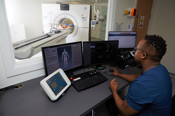
- The images are acquired as the table moves back and forth through a “donut-shaped” machine.
For most outpatient appointments, you will be able to leave immediately after your procedure and there are typically no restrictions on eating, drinking or driving following your scan. If you had an injection of contrast dye, you may be asked to wait for a short period before leaving.
Your scan will be reported by a consultant radiologist. The results will be sent directly to your referring doctor.
We are all exposed to natural background radiation every day. Medical exposures give a small additional dose on top of natural radiation.
The amount of radiation received during a CT is low, resulting in the equivalent of approximately a few months to two year’s background radiation.
The only effect on the patient that is known to be possible at these low doses is a very slight increase in the chance of cancer occurring many years or decades after the exposure.
As long as it is clearly necessary to help make the correct diagnosis and treatment decision, the benefits of detection, diagnosis and treatment resulting from the CT examination should outweigh these small radiation risks.
Opening Hours - Dublin Locations
Monday – Friday: 8am - 8pm
Saturday - Sunday: 8am - 5pm
Dublin Pricing
CT from €525 | Coronary CT €735
If you have private health insurance please check with your provider to find out if you have full or partial cover for this scan. Please note you might need to pay at the time of your appointment and submit a claim to your insurance provider afterwards. For more information visit our Insurance Cover page.
New Cardiac CT Scanner at Mater Private Network, Cork
Mater Private Network in Cork is one of the first hospitals in Ireland to use a new Cardiac CT scanner that is designed for use exclusively with cardiology patients. It uses the latest state-of-the-art technology to achieve superior image quality, capturing the full heart on one image and in just one beat. The aorta is imaged in less than seven seconds, and the shorter scan times ensure the lowest possible radiation exposure for your patients. The wide-bore scanner is also a more comfortable experience, especially for patients who find it difficult in confined spaces.
Services available include fast access to CT Angiogram and CT Cardiac Calcium Scoring. All scans will be reported on by a consultant who holds a sub-specialist fellowship in cardiac imaging.
Frequently asked questions
CT scanners are much quieter than an MRI. There is some noise, but it is not very loud.
A CT scan will be recommended based on the information and images that your doctor requires to make a diagnosis, a treatment plan or to assess any change in a condition. Your doctor will be able to answer this question.